Preparing your experience, please wait…

K
Home Spot DxSubmit a Spot Dx


Spot Dx
Clinical reasoning prompts
Brief cases to sharpen your diagnostic reasoning. Think through the question, reveal the answer, and join the WhatsApp discussion.
Submit your own Spot Dx
Share a diagnostic case and earn 100 XP + 40 points when published.
Cardiology
14-year-old male with sudden left-sided weakness
A 14 years old boy is referred with sudden-onset left-sided hemiparesis that began 6 hours ago. His mother reports a 3-year history of progressive exertional dyspnea and easy fatigability, for which he was repeatedly treated with salbutamol inhalers at a local clinic under a diagnosis of refractory asthma. Two weeks ago, he had a mild diarrheal illness and has since been poorly hydrated. On examination, he is afebrile but has marked central cyanosis and Grade 3 digital clubbing. Oxygen saturation is 76% on room air, rising only to 79% on high-flow oxygen. Neurological exam confirms left-sided hemiplegia (power 1/5). Cardiovascular exam reveals a prominent left parasternal heave and a palpable, loud, single second heart sound (P2). No murmurs are audible. Complete blood count shows a hemoglobin of 21.8 g/dL and a hematocrit of 66%. Mild conjunctival icterus is noted.
In cyanotic congenital heart disease, the disappearance of a murmur is a sign of clinical worsening (Eisenmenger syndrome), not improvement. Never perform phlebotomy for reactive polycythemia in a dehydrated patient, as this precipitously increases the risk of stroke; prioritize volume-for-volume fluid resuscitation first.
Nurse
Nurse
45-year-old female Immunosuppressed (ISS) on ART (likely HIV-positive)
A 45-year-old female on antiretroviral therapy (ART) with a 2-month history of progressive bilateral lower limb weakness. She presents with spastic paraparesis (power 2/5) and preserved sensorium. The pelvic X-ray shows destructive changes in the lumbosacral spine. She is immunosuppressed, likely due to HIV/AIDS. The patient has been experiencing worsening symptoms over the past two months, including difficulty walking and maintaining balance. She has not had any recent trauma or infections. Her current medications include ART, and she has been adherent to her regimen. Physical examination reveals bilateral lower limb weakness, with a power of 2/5. The patient's sensory examination is normal. Laboratory results show a low CD4 count of 150 cells/μL.
In a patient with HIV/AIDS and progressive bilateral lower limb weakness, spinal TB should be considered high in the differential diagnosis, especially when there are radiographic findings of vertebral destruction. Early initiation of anti-TB therapy is crucial to prevent further neurological deterioration.
Obstetrics
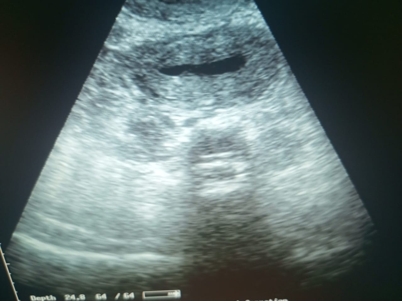
Mild lower abdominal pain 3 weeks post C-section
Mild lower abdominal pain 3 weeks post C-section No fever No pv discharge
Orthopedics
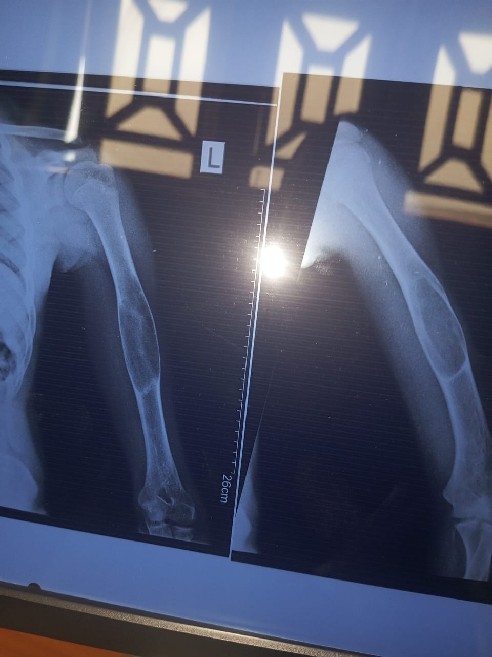
Teenager with arm swelling
13 year with swelling of left upper arm for 4 months. Has developed significant pain 3 days ago.
Infectious Diseases
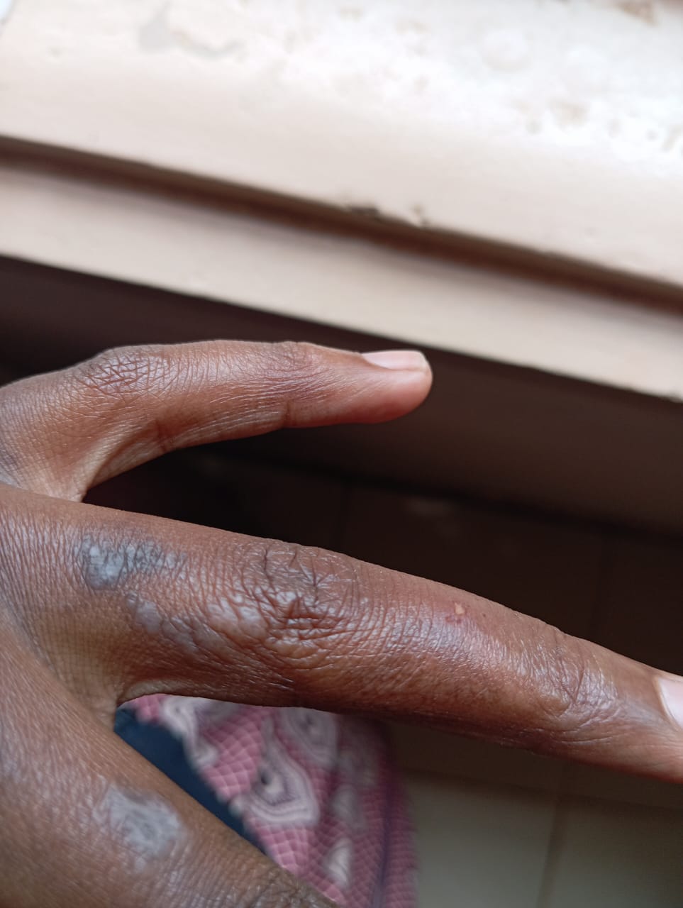
17/F with rash
A 17 y/f has heard this rash - recurrent, on and for a period of 2 years. Resolves on medication but re-occurs after like 3 weeks. It appears on hands and thighs. Currently on menthotrexate and prednisolone.
Infectious Diseases
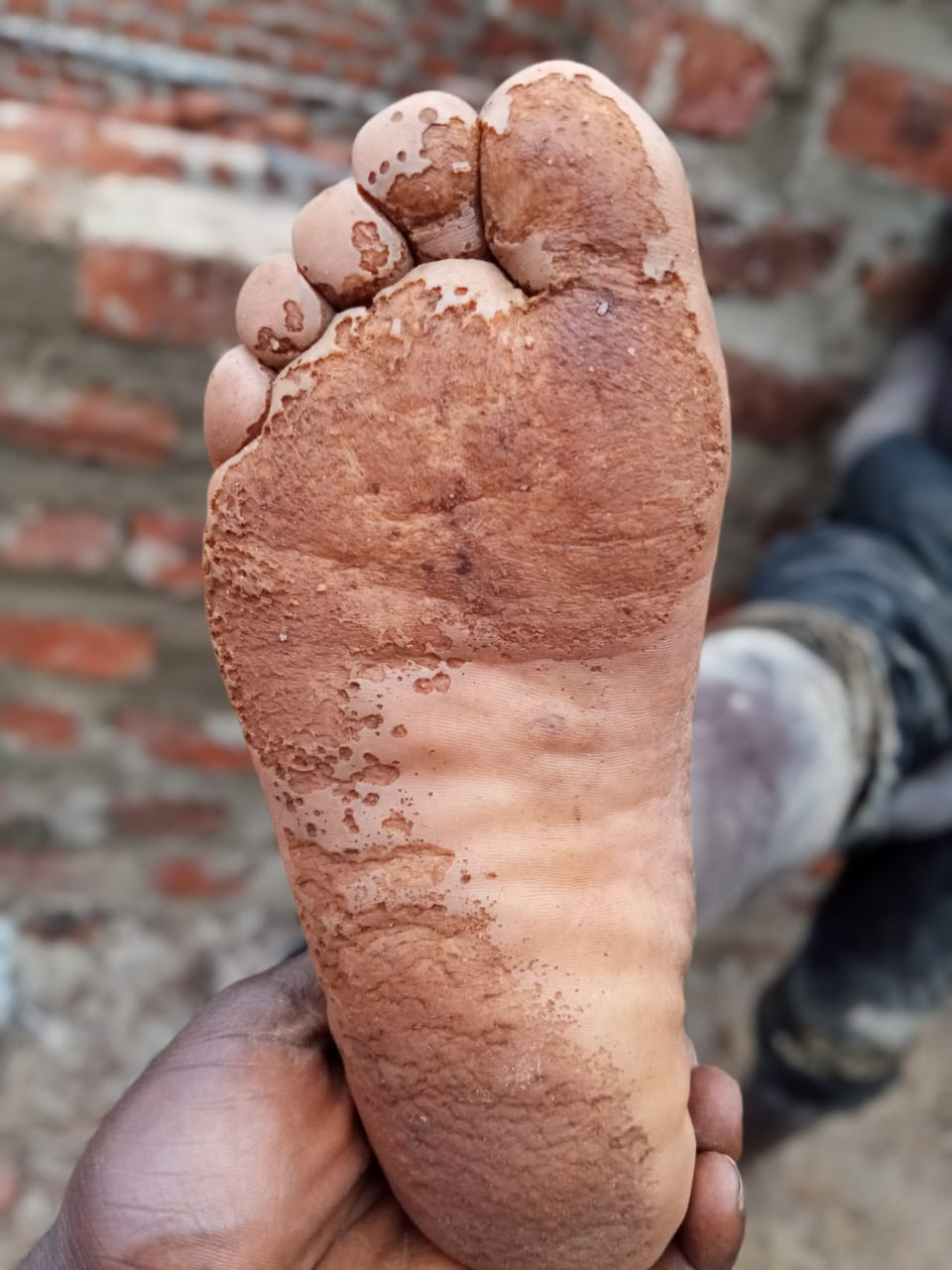
Pitting Plantar lesions
Pitting Plantar lesions
Pulmonology
68/M with ARDS
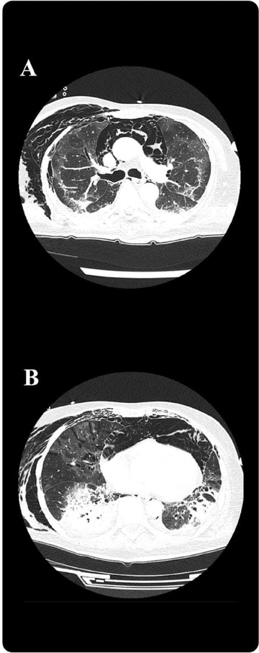
A 68-year-old man with influenza-associated acute respiratory distress syndrome developed subcutaneous emphysema and pneumomediastinum 7 days after symptom onset while receiving high-flow nasal cannula therapy. Venovenous extracorporeal membrane oxygenation (VV-ECMO) was initiated, followed by lung-protective mechanical ventilation. Bronchoscopy showed no major tracheobronchial injury. On day 11, the patient developed impaired venous drainage and hypotension (systolic blood pressure <60 mmHg), requiring norepinephrine and vasopressin. Physical examination showed deteriorated palpable crepitus in the chest and neck. Chest computed tomography was performed to evaluate the cause of refractory hypotension, impaired ECMO drainage, and extensive subcutaneous emphysema.
Infectious Diseases
45/F ISS with lower limb weakness
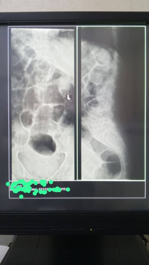
45/F ISS on ART with 2/12 hx of progressive bilateral lower limb weakness, unable to walk unaided now. Markedly reduced muscle power in both limbs (2/5), spastic with preserved sensorium.
Infectious Diseases
40/M ISS with waterly stools
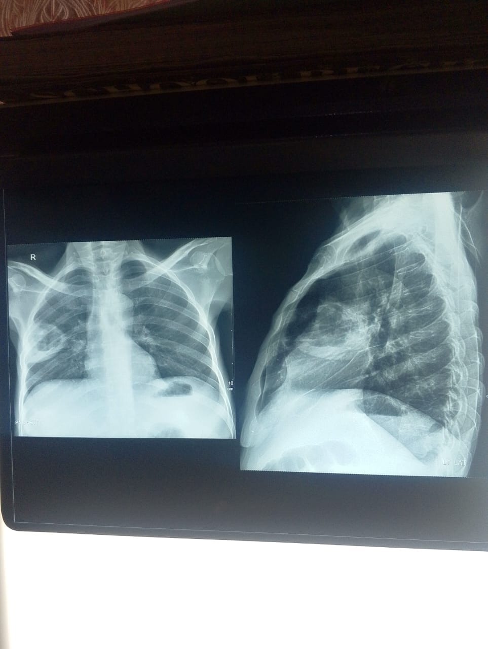
40 year old male, known ISS, on 3TC, TDF, EFV, adherence not ascertained, admitted with waterly stools, non bloody, reduced appetite, vomiting, general body weakness, cough not responding to azithromycin, Sodium: 130.7mmol/l, Potassium: 2.58mmol/l CBC - unremarkable except Hb: 5.7g/dl Urine lam- negative Modified ZN- no oocytes seen. Stool microscopy - unremarkable. Chest X-ray is attached
6/M with skin peeling
6years/male with 3 day history of this peeling off of the skin. Caretaker reports it started peeling off from the face now entire body. No use of any unusual cosmetic
Paediatrics
10/M with one week history of dry cough.
10/M with one week history of dry cough has been on Ceftriaxone-salbactam, Gentamicin, Azithromycin and prednisone for Pneumonia in a peripheral facility then referred with worsened difficulty in breathing 3 hours prior.
Internal Medicine
15/F with continuous rhythmic shaking
15/F with no known chronic illnesses presented with generalized rhythmic body shaking
Dermatology
40/M with dry scaly skin
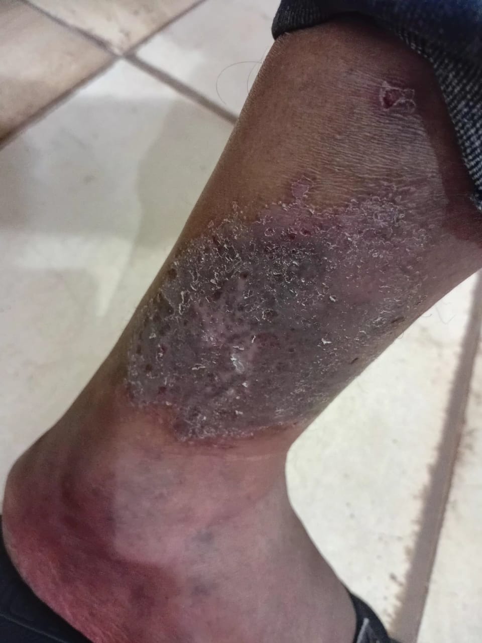
40/M with no known chronic illness presented with dry scaly skin patch on the lateral aspect of the right leg
Obstetrics & Gynecology
Obstetrics & Gynecology
24/F with acute lower abdominal pain, mild fever, and shock
A 24-year-old female is rushed to a Health Center IV in Eastern Uganda with severe, acute-onset lower abdominal pain of 6 hours, which started in the right iliac fossa and is now generalized, radiating to her right shoulder. She collapsed once at home. She reports her last menstrual period was 3 weeks ago and was "normal," though she has experienced mild, foul-smelling vaginal discharge for two weeks, for which she self-medicated with oral amoxicillin. On examination, she is pale, cold, and clammy. Axillary temperature is 37.9°C, blood pressure is 82/50 mmHg, heart rate is 132 bpm (weak), and respiratory rate is 26 breaths/minute. Her abdomen is distended, rigid, with generalized tenderness and rebound guarding. Pelvic examination reveals cervical motion tenderness and a boggy fullness in the pouch of Douglas. A bedside urine pregnancy test is faintly positive.
Do not rule out ectopic pregnancy based on a 'recent normal period' or mild fever; decidual bleeding mimics menses, and hemoperitoneum causes chemical peritonitis with reactive pyrexia. Perform culdocentesis to confirm blood, and avoid aggressive fluid resuscitation (keep SBP 80-90 mmHg) to prevent dislodging the life-saving clot before laparotomy.
42/F known ISS on ART with severe anaemia
42/F known ISS on ART for 4 years with severe anemia, hematemesis, hematochezia plus melena stools. She had similar episodes before with h/o transfusion, and has had the oral lesions for more than 20 years. No other skin lesions O/E: wasted, severe parlor, Jº, Eº, Lº kiolonychia PA: diffuse tenderness more in the epigastrium, no masses palpable Reported one of family members with similar lesions No Family hx of cancer.
Urology
Urology
16/M with severe lower abdominal pain.
A 16-year-old boy is brought from school with sudden severe lower abdominal pain, worse toward the left groin, and two episodes of vomiting. He has no fever, dysuria, urethral discharge, or diarrhoea. He had been receiving first aid for bruises after a fight with a classmate, but the pain remained severe despite analgesia. On examination, he is uncomfortable, with mild left iliac fossa guarding and marked left groin tenderness. No obvious groin swelling is seen. The patient's symptoms have been ongoing for several hours, and he has been unable to urinate since the onset of pain.
In cases of suspected testicular torsion, do not delay surgical consultation to obtain an ultrasound or other imaging; the diagnosis is primarily clinical, and timely surgical intervention is critical to salvage the affected testis.
Endocrinology
Endocrinology
9/M with rapid deep breathing, confused, and lethargic
A 9-year-old boy is brought to the emergency department in a confused and lethargic state. His mother reports that he has been excessively thirsty and passing large amounts of urine for the last two weeks, and has recently started vomiting. On examination, he is severely dehydrated with a respiratory rate of 35/min, and his breathing is deep and rhythmic.
In any child presenting with rapid, deep breathing (air hunger) and signs of dehydration without a clear respiratory cause, check the blood glucose to exclude DKA.
Cardiology
Cardiology
65/M with new heart murmur, fever, and small spots on palms
A 65-year-old male presents with a persistent low-grade fever and weight loss over the last 4 weeks. He mentions a history of a dental extraction two months ago. On examination, a new pansystolic murmur is heard at the apex, and small, non-tender, reddish spots are seen on his palms (Janeway lesions). He also has tiny 'splinter-like' hemorrhages under his fingernails.
The presence of a new heart murmur in a patient with unexplained fever should be considered infective endocarditis until proven otherwise; look for stigmata like Janeway lesions and splinter hemorrhages.
Nephrology
Nephrology
60/F with 3-month tiredness, pallor, and high blood pressure
A 60-year-old female presents with a 3-month history of progressive tiredness and generalized itching. She has a long-standing history of hypertension which she manages inconsistently with herbal remedies. On examination, she has marked conjunctival pallor and her blood pressure is 175/105 mmHg. Laboratory tests reveal a hemoglobin of 8.2 g/dL, a serum creatinine of 450 µmol/L (4.9 mg/dL), and proteinuria 3+ on dipstick.
Chronic Kidney Disease is a silent killer; normochromic normocytic anemia and resistant hypertension in a middle-aged patient are hallmark signs of advanced renal parenchymal disease.
Nutrition
Nutrition
2/F with swelling of limbs, skin peeling, and 'miserable' mood
A 2-year-old girl is brought to a health center because her legs and face have become swollen over the last month. Her mother reports she is 'always crying' and has lost her appetite for food. On examination, she has pitting edema up to her knees, thin reddish hair, and dark skin lesions on her thighs. Her heart rate is 85 bpm and her temperature is 35.5°C.
In a child with SAM, standard vital sign thresholds for infection (like tachycardia or fever) may be absent; bradycardia and hypothermia are late indicators of severe sepsis or electrolyte derangement.
Cardiology
45/F with acute chest pain and dyspnea
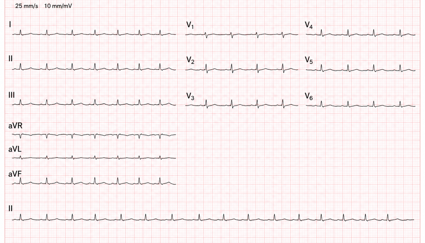
A 45-year-old female, who had recently been evaluated at a local clinic for symptoms of a febrile illness, presents to the emergency department with acute shortness of breath and chest pain that worsens with lying down. She reports a 2-day history of fever and dry cough, but no one with similar symptoms at home. Clinical examination reveals a blood pressure of 90/60 mmHg, a heart rate of 115 bpm, and an elevated jugular venous pressure (JVP). Heart sounds (1 & 2) are heard, but distant. An ECG shows low voltage QRS complexes, but no obvious ischemic changes. Troponin levels are mildly elevated, but not significantly so for her presentation. She has no history of heart disease, but her family reports a delay in seeking medical care due to financial constraints.
In resource-limited settings, bedside ultrasound can be a valuable tool for rapid diagnosis of life-threatening conditions like cardiac tamponade, allowing for timely intervention and potentially improving patient outcomes. However, as it is not always available, high index of suspicion should call for early escalation if unable.
Internal Medicine
Cough and weight loss in a 34-year-old farmer
A 34-year-old tobacco farmer from Mukono presents with a 6-week history of productive cough, night sweats, and unintentional 8 kg weight loss. He has no prior medical history. On examination: BMI 17.2, bilateral coarse crackles in the upper zones, no peripheral lymphadenopathy.
Upper-zone crackles + constitutional symptoms in a high-burden setting = think TB first, not pneumonia.